KMC tops in imaging field

A cure for cancer is still wishful thinking, but Inland Northwest residents have access to the next best thing.
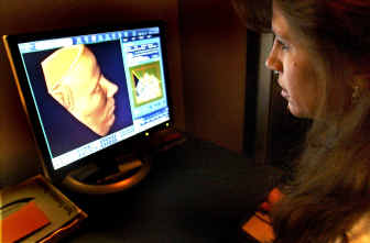
“This is as close to the magic bullet phenomenon as we can get,” said Dr. Arne Michalson, a radiologist and director of nuclear medicine at Kootenai Medical Center in Coeur d’Alene, as he studied a three-dimensional image of a patient’s esophagus on his computer screen. “What we’re trying to do is find, characterize and treat problems earlier with less radiation, less overall cost, less morbidity.”
During the last three years, KMC has built itself into one of the nation’s 101 most-wired hospitals with its radiology information system, according to the American Hospital Association. Its high-tech medical imaging equipment set the standard in the Inland Northwest for a short time. Such technology is now available in Spokane and Boise, but rare outside most metropolitan areas in the country. Only Stanford University beat KMC in the West using the newest high-tech CT scan available.
Medical images like X-rays and MRIs are revealing patients’ internal abnormalities, such as beginning tumors, earlier than ever before, leading to early and more effective treatment and, ultimately, longer lives.
X-rays and other medical tests doctors order at KMC for their patients no longer include film or light stations to read the film. Instead, high-tech scanners take pictures of patients’ insides with 10 times the detail and much less potential for error than film. Computer programs enable radiologists to view the images from all angles in a variety of shadings and sizes that help emphasize irregularities. Budding tumors that often don’t show up on film appear in the high-tech images.
Technology makes those images available through a secure network to doctors at home and hospitals in other areas. Radiologists consult with specialists, other radiologists or a patient’s doctor at any distance via computer and view the same image at the same time. Repeat MRIs or X-rays are unnecessary when patients are moved from one hospital to another.
“The faster I can diagnose based on these images and get it to the referring doctor, the faster the patient gets treatment,” Michalson says.
Five radiologists worked with KMC when Michalson, a Moscow High graduate, joined the staff in 1996. Filmed images of patients’ bones or organs were delivered to them by hand. They plugged the film into light boxes and recorded their observations onto cassettes. Their notes were transcribed and sent to the doctors who ordered the images.
“It took up to two days and that’s how it’s still done in most hospitals,” says Dr. Scott Venera, KMC’s director of radiology.
New technology spotlighted how slow the film process was.
“We wanted to be ahead of the curve, embrace the technology and go with it,” Michalson says.
The price didn’t deter Michalson and Venera. They needed two special labs at $1.5 million each and a $3 million imaging center. They needed two high-tech MRI machines at $2.2 million each and two CAT scan machines at $1.6 million each. Two more special scanners called PET CTs cost $2.5 million each.
The group of radiologists, North Idaho Imaging, worked as partners with KMC. The hospital proposed the arrangement years ago to avoid competition with a for-profit group. Working as partners eliminated the need for separate equipment and staffs, says Tom Legel, KMC’s chief financial officer.
Michalson and Venera proposed the $18 million upgrade in medical imaging to KMC and trustees didn’t blanch.
“We try to anticipate replacement or the addition of new equipment,” Legel says. “New equipment generates new revenue. We also try not to buy everything in one year. We stagger the purchases.”
Equipment wasn’t the only cost. Radiologists and MRI and X-ray technologists needed new training. The high-tech machines produced wafer-thin – .75 mm thick – images of body parts. Radiologists began seeing more than they’d ever seen. The technology offered views that hadn’t been possible.
Janet Dorendorf, KMC’s MRI supervisor, learned to ask patients their symptoms in case they told her something they’d forgotten to tell their doctors. So many image options are available that Dorendorf personalizes patients’ pictures as much as possible.
“That’s the art,” she says. “We can tailor the exam to the needs of the patient.”
CT/MRI technologist Leah Byrd traveled the world to learn the latest in imaging. She can perform a virtual colonoscopy on her computer screen with an image of a patient’s colon. Seated at her desk, Byrd opens an image of the colon from the inside. She manipulates her mouse so she appears to travel through the colon as if she were in a tunnel. The image shows her every ripple, fold and flaw. Byrd marks abnormalities to help radiologists with their diagnoses.
Despite the cost, radiology information systems are slowly spreading to larger hospitals nationwide. Insurance providers pay the same for images from the new technology as they do for images on film, KMC’s radiologists say.
KMC became completely film-free in December. Patients’ images are stored in computer files now, easily accessed and sent anywhere in seconds. Michalson grins like a little boy on Christmas morning as he sits in front of five computer screens revealing cracks in bones, collapsing arteries and tumors the size of a pinhead.
“Radiologists are people. Hey, that could be me,” he says. “We want to provide the kind of care we’d want for ourselves. With this product, we went from a box ukulele to a Stradivarius.”